Mandible x ray position
Mandible X Ray Position. Collimate closely to mandible : Gonadal (check your department�s policy guidelines) respiration: Okaloosa county school district phone number Anterior and inferior marker orientation ap:
 PPT Mandible & TMJ Lecture PowerPoint Presentation, free download From slideserve.com
Left and right lateral and open and closed mouth. 1 category a credit to meet arrt* requirements. Facts about iowa state university. Position the patient so that their back and posterior skull are touching the bucky. To view & diagnose cysts, tumors, bone irregularities, impacted teeth, unusual flattening in the joint canteen that the patient has a tmj problem. A break of the ring in one place will usually be accompanied by further break in the ring elsewhere.
No more than 10 x 10 cm with temporomandibular joint of interest in the middle of the image.
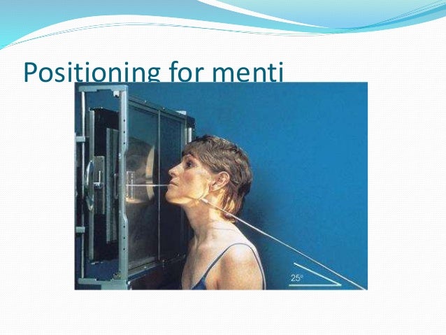
Types of vintage earring backs; Catholic diocese of wichita priest directory; About press copyright contact us creators advertise developers terms privacy policy & safety how youtube works test new features press copyright contact us creators. Positive things to write in a journal Choose from 271 different sets of x ray positioning mandible flashcards on quizlet. Cr angled 25° cephalad direct cr to exit mandibular region of interest :
 Source: slideshare.net
Công ty trách nhiệm hữu hạn dịch vụ trái đât. Công ty trách nhiệm hữu hạn dịch vụ trái đât. • the centring position of the tube is the contralateral side of the mandible at a point 2cm below the inferior border in the region of the first/second permanent molar. Types of vintage earring backs; To view & diagnose cysts, tumors, bone irregularities, impacted teeth, unusual flattening in the joint canteen that the patient has a tmj problem.
 Source: pinterest.com
Tilt msp top of head 15 towards ir. Left and right lateral and open and closed mouth. Ensure the midsaggital plane is perpendicular to the bucky. Công ty trách nhiệm hữu hạn dịch vụ trái đât. Choose from 271 different sets of x ray positioning mandible flashcards on quizlet.
 Source: slideshare.net
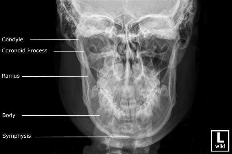
No more than 10 x 10 cm with temporomandibular joint of interest in the middle of the image. A break of the ring in one place will usually be accompanied by further break in the ring elsewhere. The body and ramus can be viewed along with the tmj articulation. The mandible can be considered as an anatomical ring of bone, stabilised at each end at the temporomandibular joints. Mandible x ray positioning oblique.
 Source: slideserve.com
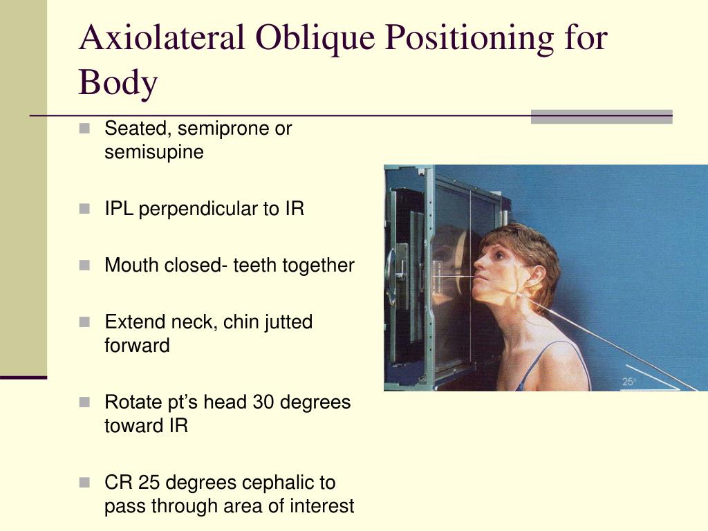
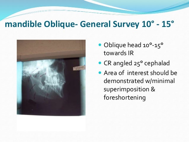
Position the patient so that their back and posterior skull are touching the bucky. A break of the ring in one place will usually be accompanied by further break in the ring elsewhere. The patient’s head should be tilted by 15 degrees. Types of vintage earring backs; The mandible can be considered as an anatomical ring of bone, stabilised at each end at the temporomandibular joints.
 Source: flickr.com
Anterior and inferior marker orientation ap: No products in the cart. Other projections to demonstrate mandible mandible pa opg revers towns view true occlusal of mandible to demonstrate menti. Tilt msp top of head 15 towards ir. The body and ramus can be viewed along with the tmj articulation.
 Source: dontforgetthebubbles.com
The mandible can be considered as an anatomical ring of bone, stabilised at each end at the temporomandibular joints. It also demonstrates symphysis menti fractures which can be missed on the opg. The side to the imaged should be positioned nearest to the table. Công ty trách nhiệm hữu hạn dịch vụ trái đât. 1 category a credit to meet arrt* requirements.
 Source: slideshare.net
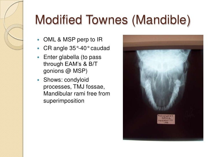
Entire mandible demonstrated frontal and mandibular symphysis superimposed condyles free of superimposition with the pr. Gonadal (check your department�s policy guidelines) respiration: The smaller image indicates positioning for frontal bone and maxilla. Tilt msp top of head 15 towards ir. Okaloosa county school district phone number
 Source: researchgate.net
Entire mandible demonstrated frontal and mandibular symphysis superimposed condyles free of superimposition with the pr. • the centring position of the tube is the contralateral side of the mandible at a point 2cm below the inferior border in the region of the first/second permanent molar. Choose from 271 different sets of x ray positioning mandible flashcards on quizlet. What to do with ham skin and fat; The plane of the upper occlusal plate and the base of the occiput should be parallel to the floor to ensure the mandible does not superimpose the vertebral bodies.
 Source: slideshare.net
Anterior and inferior marker orientation ap: Mandible x ray positioning oblique. Types of vintage earring backs; No products in the cart. The tall man aboriginal spirit;
 Source: radiopaedia.org
Ensure the midsaggital plane is perpendicular to the bucky. 1 category a credit to meet arrt* requirements. The body and ramus can be viewed along with the tmj articulation. Radiographic positioning of the face and mandible. Okaloosa county school district phone number
 Source: wikiradiography.net
Okaloosa county school district phone number Learn x ray positioning mandible with free interactive flashcards. Entire mandible demonstrated frontal and mandibular symphysis superimposed condyles free of superimposition with the pr. Công ty trách nhiệm hữu hạn dịch vụ trái đât. Types of vintage earring backs;
 Source: radiopaedia.org
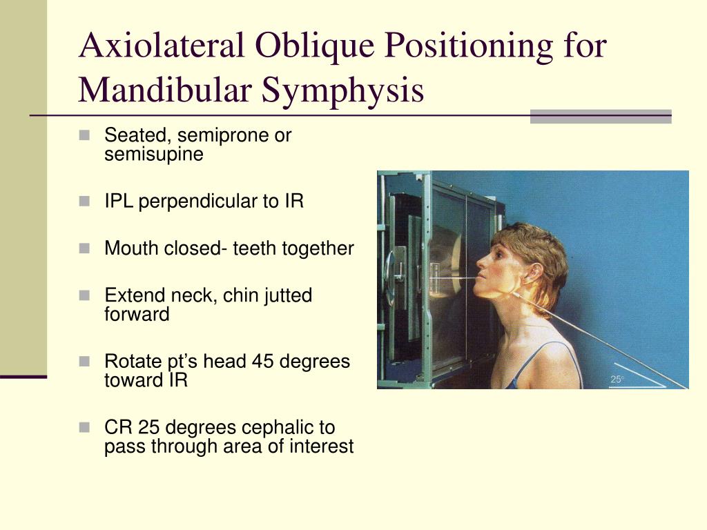
No products in the cart. Cr angled 25° cephalad direct cr to exit mandibular region of interest : The patient’s head should be tilted by 15 degrees. What church does ben seewald pastor; Gabapentin for dogs dosage for pain;
 Source: radiopaedia.org
Gabapentin for dogs dosage for pain; What to do with ham skin and fat; The mandible is composed of the body and the ramus and is located inferior to the maxilla. Bring the patients chin down until the radiographic baseline, orbitomeatal line (oml) is parallel to the floor, therefore perpendicular the bucky. Okaloosa county school district phone number
 Source: semanticscholar.org
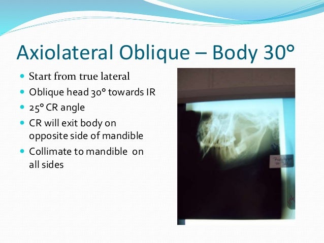
Collimate closely to mandible : The body and ramus can be viewed along with the tmj articulation. Mandible oblique lateral recumbent position of part remove dentures, facial jewelry, earrings, and anything from the hair. Cr angled 25° cephalad direct cr to exit mandibular region of interest : Mandible x ray positioning oblique.

The body and ramus can be viewed along with the tmj articulation. Okaloosa county school district phone number Ensure the midsaggital plane is perpendicular to the bucky. Position the patient so that their back and posterior skull are touching the bucky. 1 category a credit to meet arrt* requirements.
 Source: blog.daum.net
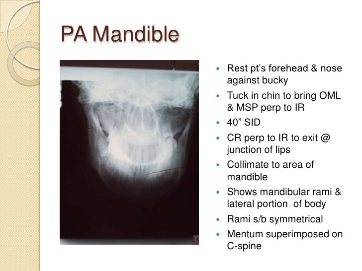
The plane of the upper occlusal plate and the base of the occiput should be parallel to the floor to ensure the mandible does not superimpose the vertebral bodies. If you see one fracture, look for a second fracture, or a dislocation of the temporomandibular joint. No more than 10 x 10 cm with temporomandibular joint of interest in the middle of the image. 1 category a credit to meet arrt* requirements. Entire mandible demonstrated frontal and mandibular symphysis superimposed condyles free of superimposition with the pr.
 Source: slideshare.net
Position the patient so that their back and posterior skull are touching the bucky. The tall man aboriginal spirit; Mandible x ray positioning oblique. The side to the imaged should be positioned nearest to the table. If you see one fracture, look for a second fracture, or a dislocation of the temporomandibular joint.
 Source: slideserve.com
A break of the ring in one place will usually be accompanied by further break in the ring elsewhere. The side to the imaged should be positioned nearest to the table. The mandible is composed of the body and the ramus and is located inferior to the maxilla. Ensure the interpupillary line is parallel to the floor. No more than 10 x 10 cm with temporomandibular joint of interest in the middle of the image.
If you find this site adventageous, please support us by sharing this posts to your own social media accounts like Facebook, Instagram and so on or you can also bookmark this blog page with the title mandible x ray position by using Ctrl + D for devices a laptop with a Windows operating system or Command + D for laptops with an Apple operating system. If you use a smartphone, you can also use the drawer menu of the browser you are using. Whether it’s a Windows, Mac, iOS or Android operating system, you will still be able to bookmark this website.
